Nuclear Medicine

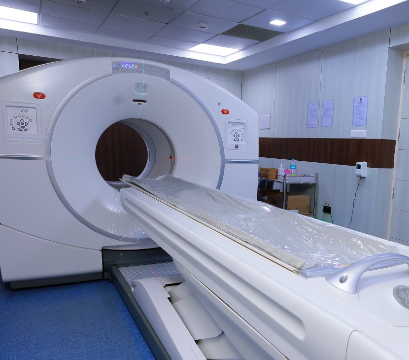
The Department of Nuclear Medicine at Thangam Hospital offers cutting-edge diagnostic imaging using Positron Emission Tomography – Computed Tomography (PET-CT) to provide early, precise detection of cancer and other serious conditions.
Our facility specializes in 18F-FDG PET scans and PSMA PET scans, helping detect, stage, and monitor various cancers with metabolic-level insights that go beyond conventional imaging.
All scans are conducted by appointment only for optimal planning, precision, and patient safety.
PET-CT Scan (Positron Emission Tomography – Computed Tomography)
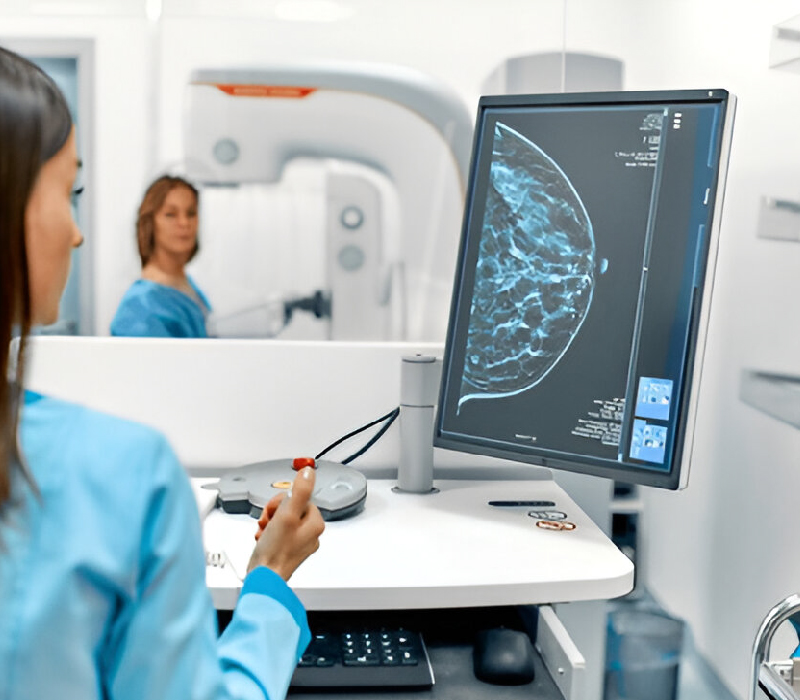
PET-CT is a powerful diagnostic tool that combines functional and anatomical imaging. It involves injecting a small amount of radioactive tracer, which accumulates in areas of high metabolic activity—such as cancer cells, infections, or inflamed tissues.
At Thangam Hospital, we perform daily 18F-FDG PET scans, where Fluorodeoxyglucose (FDG)—a radioactive glucose compound—is administered. Cancer cells absorb this tracer more rapidly, appearing as “hot spots” on the scan, helping doctors:
- Detect cancer early
- Assess the extent of spread (staging)
- Monitor treatment effectiveness
- Identify recurrence or residual disease
Applications Beyond Oncology
While PET-CT is a cornerstone in cancer imaging, it is also valuable for diagnosing other complex conditions:
- Brain Disorders
- Evaluation of brain tumors
- Detection of Alzheimer’s disease
- Localization of epileptic foci in seizure disorders
- Cardiac Imaging
- Assessment of myocardial viability
- Analysis of heart function and perfusion
- Infections and Inflammation
- Identify hidden infections or septic foci
- Evaluate autoimmune diseases and inflammatory processes
PSMA PET Scan (Prostate-Specific Membrane Antigen Imaging)
The PSMA PET-CT scan is a highly targeted imaging tool for diagnosing and managing prostate cancer. It uses a tracer that binds to the PSMA protein, which is overexpressed in prostate cancer cells. This results in high-resolution images that:
- Detect even small metastases missed by other scans
- Help guide surgical planning or radiotherapy
- Monitor treatment response and disease progression
The PSMA PET scan is particularly useful for patients with recurrent or high-risk prostate cancer.
High-Precision Imaging with SPECT/Gamma Camera for Functional Diagnosis
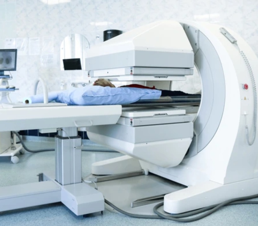
The Department of Nuclear Medicine at Thangam Hospital is equipped with advanced Single Photon Emission Computed Tomography (SPECT) and Gamma Camera technology to provide functional imaging for a range of conditions, including cancer, cardiac, renal, and bone disorders.
SPECT scans offer detailed, real-time insights into how organs and tissues are functioning, often identifying abnormalities before structural changes appear. Our Gamma Camera system ensures high-resolution imaging with low radiation exposure, enabling accurate diagnosis and effective treatment planning.
All SPECT studies are performed strictly by appointment to ensure patient-specific protocols and optimal image quality.


Theranostics: Targeted Therapy Meets Precision Diagnosis
At Thangam Hospital, our Nuclear Medicine Department offers Theranostics—an advanced, dual-purpose approach that combines targeted therapy with diagnostic imaging for highly personalized cancer care.
This technique uses radiopharmaceuticals that not only help detect cancer cells through imaging but also deliver therapeutic radiation directly to the tumor, minimizing damage to healthy tissue. It is especially effective in treating neuroendocrine tumors and metastatic prostate cancer using agents like Lutetium-177 and Actinium-225.
All Theranostic procedures are carefully planned and conducted by our expert team under strict safety protocols and are available on a scheduled, appointment-only basis.

The Department of Nuclear Medicine at Thangam Hospital offers cutting-edge diagnostic imaging and targeted radionuclide therapies, combining precision diagnosis with advanced treatment options. Equipped with state-of-the-art facilities, our team delivers personalized, safe, and effective care for cancer and other complex conditions.
Facilities
- PET-CT Scanner – High-resolution metabolic imaging for oncology, neurology, cardiology, and infection-related disorders.
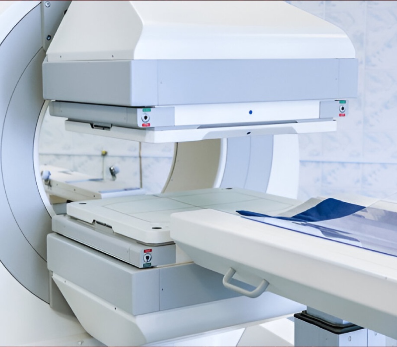
- Gamma Camera / SPECT-CT – Advanced functional imaging for organ-specific studies.
- Radionuclide Therapy Ward – Dedicated and safe setup for targeted radioisotope therapies.
PET-CT scan (Positron Emission Tomography – Computed Tomography)
PET-CT combines functional and anatomical imaging, using a small radioactive tracer that accumulates in areas of high metabolic activity such as cancer cells, infections, or inflammation.
Types of PET-CT scans available at Thangam Hospital:
- 18F-FDG PET Scan – Daily scans for detecting, staging, and monitoring various cancers; also valuable in brain, cardiac, and infection imaging.
- PSMA PET Scan – Highly sensitive imaging for prostate cancer, enabling detection of small metastases, guiding surgical and radiation planning, and monitoring treatment response.
Applications of PET-CT:
- Early cancer detection and staging
- Monitoring treatment effectiveness
- Identifying recurrence or residual disease
- Evaluating brain disorders (tumors, Alzheimer’s, epilepsy)
- Assessing cardiac viability and perfusion
- Locating hidden infections or inflammatory foci


SPECT-CT / Gamma Camera Imaging
With advanced Gamma Camera and SPECT-CT technology, our Nuclear Medicine Department provides functional diagnosis for renal, cardiac, skeletal, endocrine, pulmonary, and gastrointestinal conditions.
Services offered:
- Renal Imaging: 99mTc DTPA/LLEC Renogram, 99mTc DMSA renal cortical scan
- Hepatobiliary & GI Imaging: 99mTc HIDA scan, 99mTc Sulfur colloid reflux study, RBC scintigraphy for GI bleed, Meckel’s scan
- Endocrine Imaging: 99mTc Thyroid scan, 99mTc MIBI parathyroid scintigraphy
- Cardiac Imaging: 99mTc MIBI myocardial perfusion imaging
- Pulmonary Imaging: 99mTc MAA lung perfusion studies
- Skeletal Imaging: 99mTc MDP bone scan, lymphoscintigraphy for sentinel lymph node mapping
Radionuclide Therapy (Theranostics)
Beyond diagnosis, the Department of Nuclear Medicine offers theranostics—a dual approach that combines targeted therapy with diagnostic imaging for highly personalized cancer care.
Therapies offered include:
- Radioiodine Therapy – For hyperthyroidism and differentiated thyroid cancer
- PRRT (Peptide Receptor Radionuclide Therapy) – Targeted treatment for neuroendocrine tumors (NETs)
- Lu-PSMA Therapy – Advanced radionuclide therapy for prostate cancer

