Comprehensive
Laboratory Services
Timely support. Accurate Diagnosis.
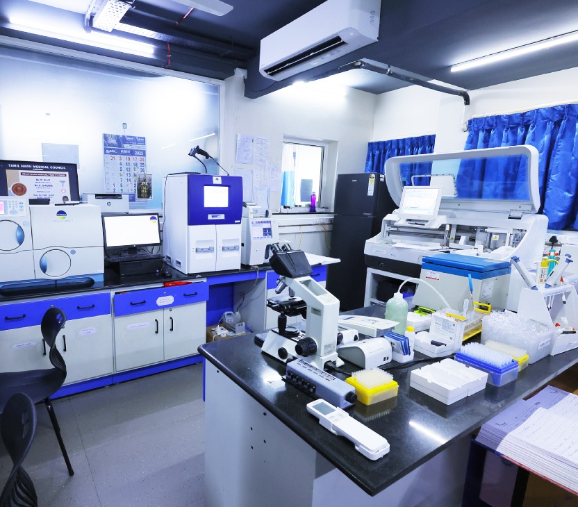
Thangam Cancer Center offers a full suite of essential laboratory services under one roof to support accurate and timely diagnosis. From routine blood tests to advanced pathology and molecular diagnostics, our in-house labs ensure high-quality, reliable results that aid in swift treatment planning and better patient outcomes.
Haematology Services
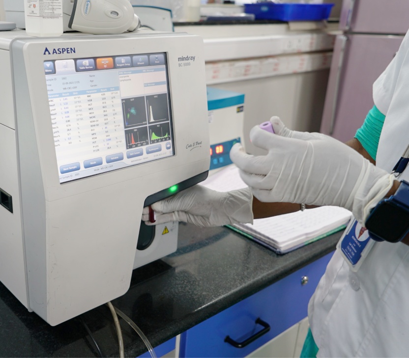
At Thangam Cancer Center, our Haematology department specializes in diagnosing and managing diseases related to blood and its components. This includes anemia, bleeding disorders, and blood cancers such as leukemia, lymphoma, and multiple myeloma. Our expertise also extends to successful bone marrow transplants, with a growing number of patients benefiting from this advanced treatment.
We are equipped with state-of-the-art diagnostic tools, including a 3-laser, 13-colour flow cytometer for detailed analysis and subtyping of blood cancers. Our lab also conducts a comprehensive range of tests to accurately diagnose anemia, clotting disorders (coagulopathies), and inherited blood conditions (hemoglobinopathies), all powered by globally benchmarked technology.
Biochemistry Services
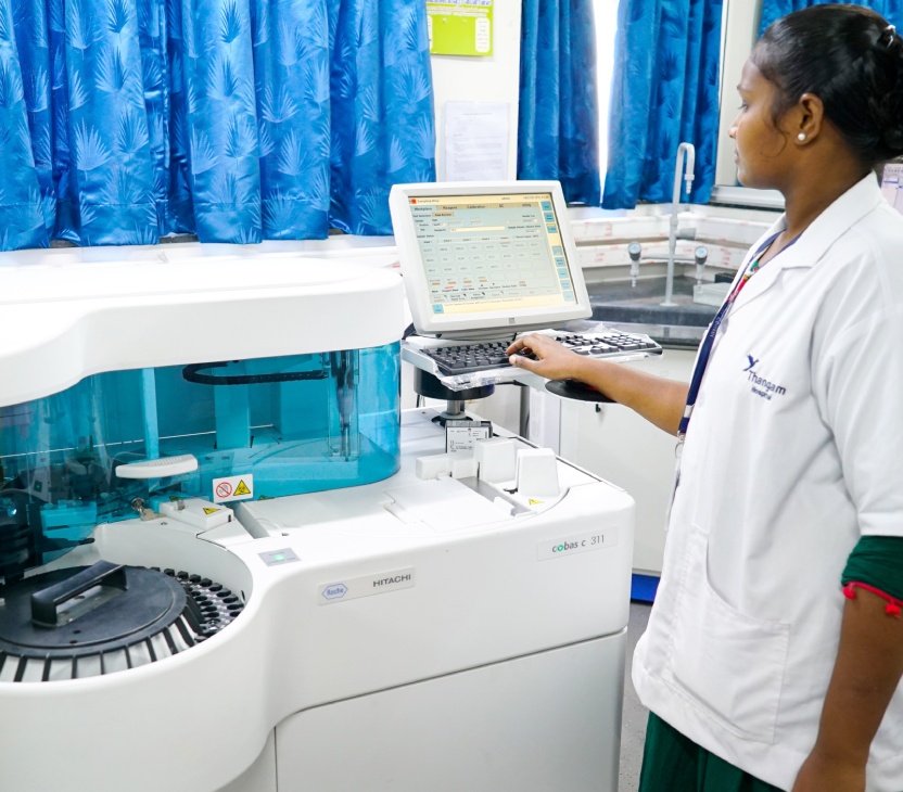
At Thangam Cancer Center, our Biochemistry lab is powered by the ROCHE COBAS c 311 analyzer, a highly advanced and versatile system that supports a wide range of clinical chemistry tests. This fully automated analyzer ensures accurate and efficient detection of critical biomarkers in various sample types including serum, plasma, urine, cerebrospinal fluid, whole blood, and hemolysate.
With over 130 test applications available, including HbA1c, electrolyte analysis (sodium, potassium, chloride), therapeutic drug monitoring (TDM), and Drug of Abuse Testing (DAT), the system offers flexibility and reliability for a wide range of diagnostic needs. Its intelligent workflow allows for continuous random access, automatic sample integrity checks, sample dilution, and reruns, all contributing to predictable turnaround times and high-quality patient care.
Immunology Services
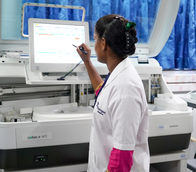
Thangam Cancer Center’s Immunology laboratory is equipped with the ROCHE cobas e 411 analyzer, a state-of-the-art system designed for high-performance immunoassay testing. Using patented ElectroChemiLuminescence (ECL) technology, this fully automated analyzer delivers precise, sensitive, and reproducible results for both quantitative and qualitative in vitro diagnostics.
The system supports a broad test menu that includes markers for anemia, bone health, cardiac function, tumor detection, fertility and hormones, maternal care, and infectious diseases. With a throughput of up to 86 tests/hour and the ability to run 18 assays simultaneously, it ensures fast and efficient processing. Emergency diagnostics benefit from 9-minute STAT testing for critical markers like Troponin T, CK-MB, Myoglobin, PTH, and hCG.
Key highlights:
- Over 100 assays available
- Error-free reagent handling with cobas e packs
- Cost-effective kit sizes with high stability and minimal wastage
- This analyzer helps us deliver timely, reliable results — a vital component in advanced cancer diagnostics and patient care.
Histopathology Services
Thangam Cancer Center’s Histopathology department is equipped with advanced technologies that ensure rapid and precise cancer diagnosis at the cellular level — even during surgery.
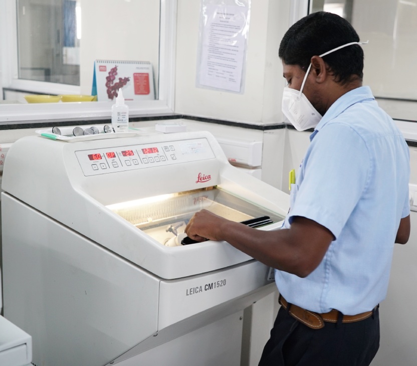
Intraoperative Diagnosis – Leica CM1520 Cryostat
With the Leica CM1520 Cryostat, we offer intraoperative consultations within 20 minutes, enabling on-the-spot decisions for procedures such as tumor margin assessment, sentinel node evaluation, and primary diagnosis. Traditionally, preparing tissue samples for microscopic analysis takes up to 48 hours. But with this system, surgeons receive critical pathological insights during surgery itself — helping improve surgical accuracy and outcomes.
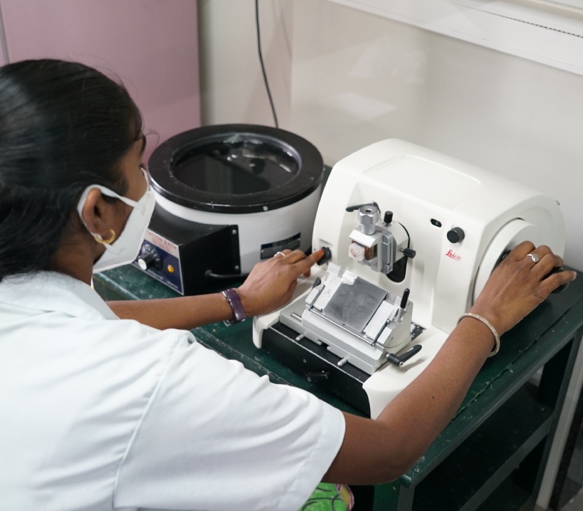
Precision Sectioning – Leica RM2125 Manual Microtome
Our Leica RM2125 rotary microtome allows highly precise sectioning, producing tissue slices as thin as 3–4 microns. This ensures high-resolution microscopic analysis, which is essential for detailed cancer diagnosis and staging.
Advanced Immunohistochemistry – Roche Ventana Platform
Our fully automated BenchMark GX Advanced Staining System from Roche enables immunohistochemistry (IHC) and in situ hybridization (ISH) for detecting specific antigens and gene expressions in tissue samples. The system automates all steps — from baking to deparaffinization to staining — delivering consistent, reliable, and reproducible results critical for identifying cancer subtypes and guiding targeted therapy.
Frozen Section (Intraoperative Consultation)
At Thangam Cancer Center, our Frozen Section service plays a critical role in delivering rapid, intraoperative diagnosis during surgeries. This specialized pathological procedure allows our team to examine tissue samples and provide a microscopic diagnosis within just 20 minutes—while the surgery is still in progress.
Traditionally, tissue processing for diagnosis takes about 24 hours, involving formalin fixation, chemical reagents, paraffin embedding, sectioning, and staining. However, with frozen section, the tissue is immediately frozen, sectioned using a cryostat, and stained — bypassing the longer routine process.
This real-time diagnosis helps surgeons make informed decisions on the operating table, especially in assessing tumor margins, sentinel nodes, and disease staging, ensuring precision and confidence during critical cancer surgeries.
Molecular Lab
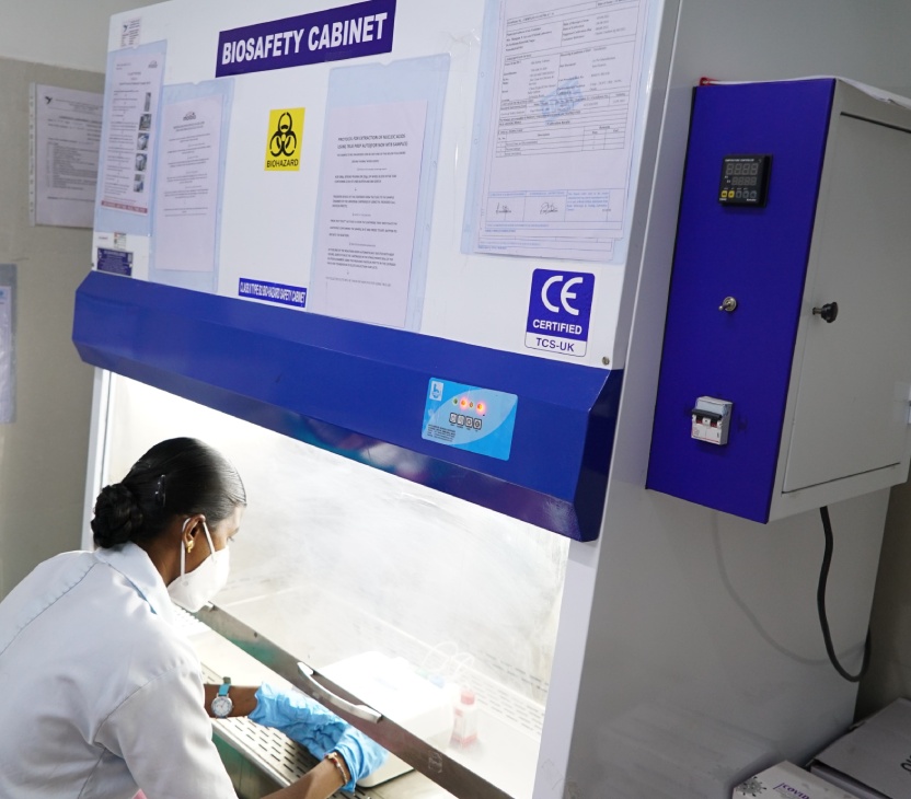
Thangam Cancer Center houses an advanced Molecular Diagnostics Lab equipped with a closed system RT-PCR platform. This cutting-edge technology enables accurate and rapid detection of infectious diseases and plays a pivotal role in the molecular subtyping of cancers.
By analyzing genetic material directly from the patient’s sample, our molecular lab helps in identifying specific mutations, gene rearrangements, and other molecular markers crucial for personalized cancer treatment. This enhances diagnostic precision, supports early detection, and guides targeted therapies—making it a vital component of our precision oncology approach.
