Esophagus Cancer
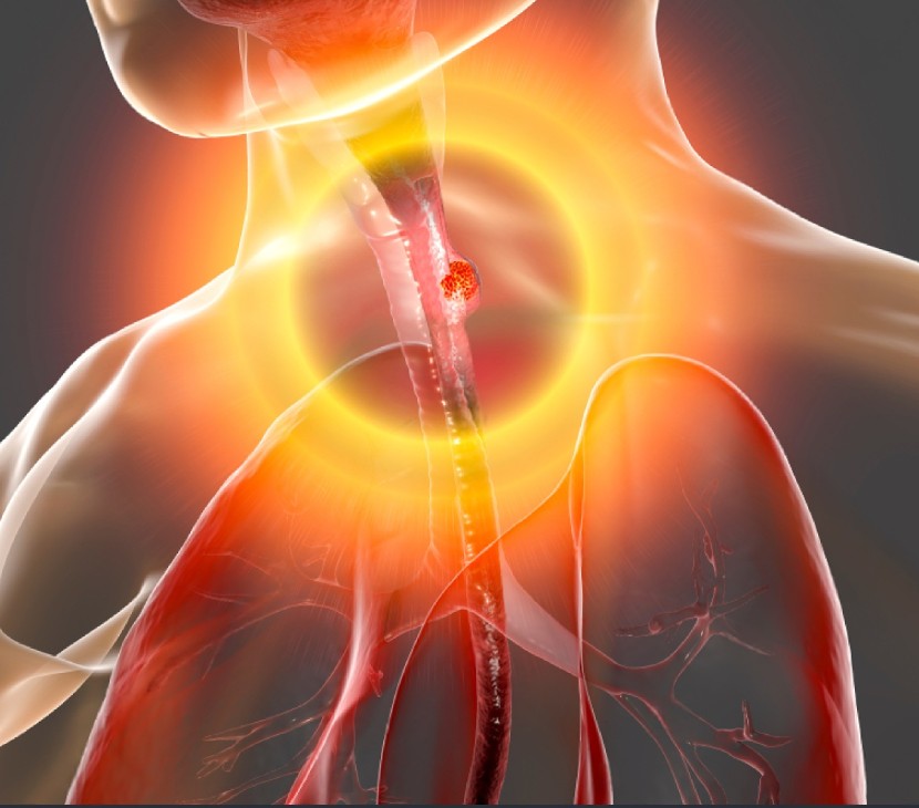
Esophageal cancer develops when abnormal cells in the food pipe develop in an uncontrolled manner. Smoking and poor acid reflux controls are major risk factors for esophageal cancer.
At Thangam Cancer Center, treatment for esophageal cancer is performed by highly trained esophageal cancer specialists resulting in systematic cancer treatment. This program provides diagnosis, recovery and supportive care for our patients
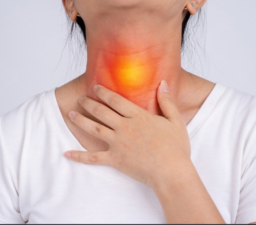
Symptoms of esophageal cancer:
People with esophageal cancer may experience subsequent symptoms or signs. Sometimes, people with esophageal cancer don’t have any of those changes. Or, the explanation for a symbol could also be a special medical condition that’s not cancer.
What is esophageal cancer?
Types of esophageal cancer
Adenocarcinoma: Adenocarcinoma starts call at glandular cells, which aren’t normally present within the lining of the oesophagus. Adeocarcinomas grow quickly due to a condition called Barrett’s esophagus, which increases a person’s chance of developing esophageal cancer. Adenocarcinoma occurs mainly at the lower end of the oesophagus and therefore the upper a part of the stomach.
Squamous Cell Carcinoma: The second commonest sort of esophageal cancer is epithelial cell carcinoma. It begins when squamous cells (thin, flat cells lining the within of the oesophagus) begin to grow uncontrollably. Epithelial cell carcinoma of the oesophagus is strongly linked with smoking and drinking an excessive amount of alcohol.
Small Cell Carcinoma: A third, rarer sort of esophageal cancer is little cell carcinoma. It begins in neuroendocrine cells, a kind of cell that releases hormones into the bloodstream in response to signals from nerves.
When to ascertain a doctor?
Most esophageal cancers do not cause symptoms in the early stages. Because symptoms typically develop once they have grown and spread, these cancers are most ordinarily diagnosed after they have advanced. Rarely are esophageal cancers diagnosed before symptoms emerge.
Screening of Cancer
The process wont to determine if cancer cells have spread within the oesophagus or to other parts of the body is named staging. It is important to understand the stage so as to plan treatment. The subsequent tests and procedures could also be utilized in the staging process:
Endoscopic ultrasound (EUS): A procedure during which an endoscope is inserted into the body, usually through the mouth. An endoscope is a thin, tube-like instrument with a light-weight and a lens for viewing. A biopsy can also be done.
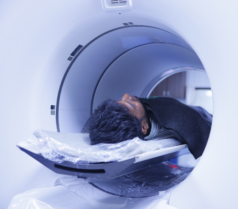
CT scan (CAT scan): A procedure that creates a series of detailed pictures of areas inside the body, like the chest, abdomen, and pelvis, taken from different angles. The photographs are made by a computer linked to an X-ray machine. In some cases, a dye is used, either injected through IV or swallowed orally, to highlight the tissue more clearly.
PET scan (positron emission tomography scan): A procedure to seek out malignant neoplasm cells within the body. A little amount of radioactive glucose (sugar) is injected into a vein. The positron emission tomography scanner rotates around the body and makes an image of where glucose is getting used within the body. A PET scan and CT scan could also be done at an equivalent time. This is often called a PET-CT.
MRI (magnetic resonance imaging): A procedure that uses a magnet, radio waves, and a computer to form a series of detailed pictures of areas inside the body. This procedure is additionally called nuclear resonance imaging (NMRI).
Thoracoscopy: A surgery to seem at the organs inside the chest to see for abnormal areas. An incision is formed between two ribs and a thoracoscope is inserted into the chest. A thoracoscope may be a thin, tube-like instrument with a light-weight and a lens for viewing. It is going to even have a tool to get rid of tissue or lymph gland samples, which are checked under a microscope for signs of cancer. In some cases, this procedure could also be wont to remove a part of the oesophagus or lung.
Laparoscopy: A surgery to seem at the organs inside the abdomen to see for signs of disease. Incisions are made on the abdominal wall and a laparoscope is inserted through them for the procedure. Other instruments could also be inserted through an equivalent or other incisions to perform procedures like removing organs or taking tissue samples to be checked under a microscope for signs of disease.
Ultrasound exam: A procedure during which high-energy sound waves (ultrasound) are bounced off internal tissues or organs, like those within the neck, and make echoes. The echoes form an image of body tissues called a sonogram. The image is often printed to be checked out later.
After esophageal cancer has been diagnosed, tests are done to seek out out if cancer cells have spread within the oesophagus or to other parts of the body.
Diagnostics Facilities
Advanced Cancer Diagnostics
Advanced Cancer Treatment
General Diagnostic Facilities
Causes
- Tobacco and alcohol: The use of tobacco products, including cigarettes, cigars, pipes, and chewing tobacco, maybe a major risk factor for esophageal cancer. The more an individual uses tobacco and therefore the longer it’s used the upper the cancer risk.
- Gastroesophageal reflux disease: People with GERD have a rather higher risk of getting adenocarcinoma of the oesophagus. This risk seems to be higher in people that have more frequent symptoms. But GERD is extremely common, and most of the people that have it don’t continue to develop esophageal cancer.
- Barrett’s oesophagus: If reflux of stomach acid into the lower oesophagus goes on for an extended time, it can damage the inner lining of the oesophagus. This causes the squamous cells that normally line the oesophagus to get replaced with gland cells. These gland cells usually appear as if the cells that line the stomach and therefore the intestine, and are more immune to stomach acid. This condition is understood as Barrett’s (or Barrett) oesophagus.
- Diet: Certain substances within the diet may increase esophageal cancer risk. For instance, there are suggestions, so far not well proven, that a diet high in processed meat may increase the prospect of developing esophageal cancer.
- High Temperatures may increase the danger for the epithelial cell sort of esophageal cancer. This could be the results of long-term damage to the cells lining the oesophagus from the recent liquids.
- Trauma: Accidentally drinking a lye-based cleaner can cause a severe chemical burn within the oesophagus. Because the injury heals, the connective tissue can cause a neighbourhood of the oesophagus to become very narrow (called a stricture). People with these strictures have an increased risk of epithelial cell esophageal cancer, which frequently occurs a few years (even decades) later.
- Plummer-Vinson syndrome: About 10% of people with this syndrome may be affected by epithelial cell cancer of the oesophagus.
Chemotherapy:
- Adjuvant chemotherapy:This type of chemotherapy is given after surgery, usually, 6 cycles with each cycle having a duration of 2-3 weeks.
- Neoadjuvant chemotherapy:This type of chemotherapy is given before surgery and is called neoadjuvant chemotherapy, consisting of 3 cycles with each cycle having a duration of 3 weeks, and, the remaining 3 cycles are given as adjuvant chemotherapy, after surgery.
- Chemo-radiation:Chemo is recommended along with radiation therapy; called concurrent chemo-radiotherapy, which can be used as a neoadjuvant or adjuvant.
- Intraperitoneal chemotherapy:This is a special form of chemotherapy delivered directly into the peritoneal (abdominal cavity) in patients with advanced stomach cancer. An intraperitoneal (IP) port system is introduced underneath the skin of the abdominal wall with a tubing passed into the abdominal cavity, under general anaesthesia. This new approach has been used successfully as neoadjuvant treatment also, especially in patients with peritoneally metastasised gastric cancer and has been shown to increase the chances of completely eliminating diseased cells with higher survival chances.
Radiation therapy:
Targeted therapy:
Immunotherapy:
Treatment:
Surgery: It is the commonest treatment for cancer of the oesophagus. A part of the oesophagus could also be removed in an operation called an esophagectomy.
Radiation therapy: Radiation therapy may be a cancer treatment that uses high-energy x-rays or other sorts of radiation to kill cancer cells or keep them from growing. The way the radiotherapy is given depends on the sort and stage of the cancer being treated. External and internal radiotherapy are wont to treat esophageal cancer. A plastic tube could also be inserted into the oesophagus to stay it open during radiotherapy. This is often called intraluminal intubation and dilation.
Chemoradiation therapy: Chemoradiation therapy combines chemotherapy and radiotherapy to extend the consequences of both.
Laser therapy: Laser therapy may be a cancer treatment that uses a beam (a narrow beam of intense light) to kill cancer cells.
Electrocoagulation: Electrocoagulation is the use of an electrical current to kill cancer cells.
Targeted therapy: Targeted therapy may be a sort of treatment that uses drugs or other substances to spot and attack specific cancer cells. Targeted therapies usually cause less harm to normal cells than chemotherapy or radiotherapy do. Antibody therapy may be a sort of targeted therapy utilized in the treatment of oesophageal cancer.
Prevention
You can take steps to scale back your risk of esophageal cancer. For instance:
Quit smoking: If you smoke, ask your doctor about strategies for quitting. Medications and counselling are available to assist you to quit. If you do not use tobacco, don’t start.
Drink alcohol carefully: If you select to drink alcohol, do so carefully. For healthy adults, meaning up to at least one drink each day for ladies and up to 2 drinks each day for men.
Eat more fruits and vegetables. Add a spread of colourful fruits and vegetables to your diet.
Maintain a healthy weight and follow an active lifestyle.